![]() Cornea Transplantation
Cornea Transplantation
What is a cornea, and what does it do?
The cornea is the clear, front part of the eye where a contact lens would sit. The cornea is responsible for two-thirds of the focusing power of the eye.
Diseases or trauma to the cornea account for about 12% of all blindness in the United States, and it is the second leading type of blindness worldwide. The cornea has five layers. All or just certain layers can be replaced with a cornea transplant. The cornea does not have a direct blood supply, meaning blood typing is not a concern in cornea transplants.
Cornea Transplantation
The transplant surgeon removes the diseased or damaged cornea from the recipient.
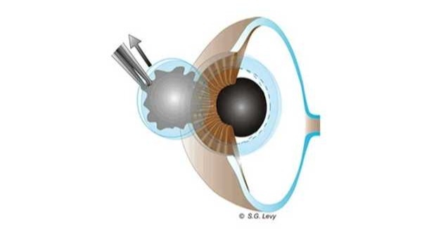
The surgeon then places a similarly-sized graft from the donor cornea into the recipient's eye. The graft is put in place by hand using microfilament sutures.
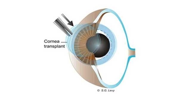
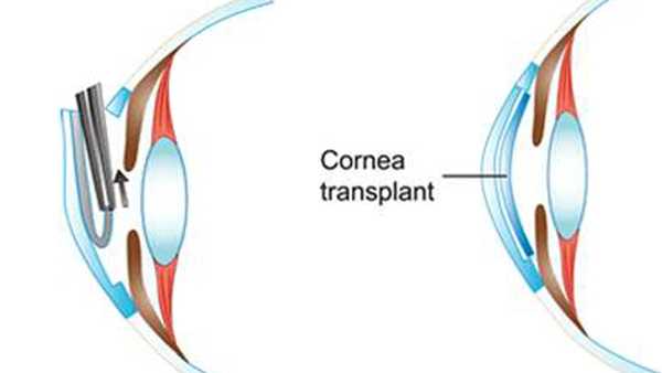
Here is the result:
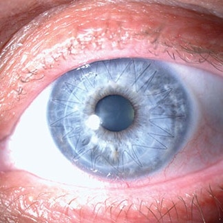
One type of procedure transplants just the endothelial layer of the cornea for certain recipients who have diseases of the endothelium. The endothelium is the deepest layer of the cornea.
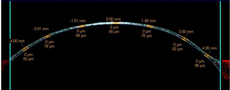
After the eye bank removes the outer portion of the donor cornea in the lab, the resulting endothelial graft is very, very thin. Here is an optical coherence tomography photo of the split graft of the donor cornea. It is only 65 microns thick. By comparison, a human hair averages 100 microns in diameter.
In surgery, the recipient's endothelium is removed through a small incision in the periphery of their cornea and the new, very thin endothelial graft is placed inside. There are no stitches and many patients see 20/20 within a few days.
If you are a cornea transplant recipient, learn more about your transplant.